Crowd-funding a high-NA multi-immersion objective lens

We have designed a promising high-NA multi-immersion all-immersion objective lens that has applications for both cleared tissue imaging and live-cell imaging.  We would love to make this objective generally available, but we need the help of the research community.

The initial design was completed just prior to the COVID pandemic and there are interested researchers, but we are stuck in a chicken-and-egg situation 1.5 years later. For custom optics like this there are large fixed costs for both design and manufacturing, so to get a reasonable per-unit price you need to make a lot of them.  But making a lot of them is a large investment that could be a loss if they don’t sell.

We were recently in a similar situation with the “King Snout” objective lens, but enough scientists placed pre-orders that ASI was comfortable proceeding with a production run. We’d like to replicate the same model with this all-immersion objective lens. Consider it a crowdfunding appeal; ASI is willing to bear significant financial risk, but only if there are enough customers who agree to purchase the resulting product.

So here’s the offer: ASI will proceed with a production run if we receive POs for seven of these objectives by end-July 2021, with expected delivery in November 2021.  We will offer a special price of $25k for any orders received by July 31. Update: We did not meet our goal of getting commitments for 7 of these, however the interested leading scientists are still interested so we are keeping a lookout for anyone else who would be willing to pre-order these.Leading scientists have already promised to purchase 3 of them, so we need another 4 customers to get us over the hump.  Any production run will be for at least 15 objectives, so there will be some left for future orders.  But the scientists who pre-order will not only be the first to get their hands on them, but will also further the entire field by helping to make this objective lens a reality.

We are happy to share CAD and Zemax black box models.  Please contact me at jon@asiimaging.com for more information or to request a formal quote.

Here are the main specifications:

  • Magnification / NA in BABB : “56x” / NA 1.24
  • Magnification / NA in CLARITY: “52x” / NA 1.15
  • Magnification / NA in water: “48x” / NA 1.05
  • Working distance: 3.4 mm
  • Optical / mechanical angle: 52° / 57°
  • Field of view: 0.5 mm
  • AR coatings: 400 – 1300 nm
  • Plan-corrected, apochromatic, optimized for visible
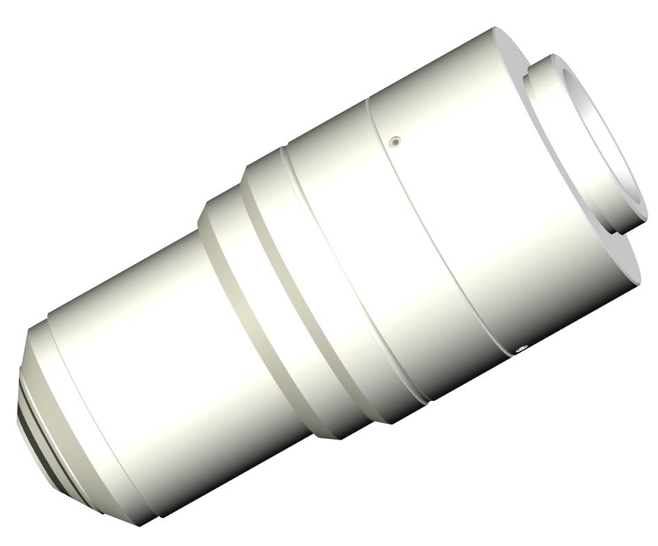
  • 83 mm parfocal distance, M25x0.75 threads

What will this objective be good for? I’m glad you asked! There are at least 3 categories of applications:

    1. Cleared tissue imaging
    2. Single-objective light sheet
    3. High-NA light sheet (e.g. lattice light sheet)

Obviously this objective lens will be an excellent choice for any application involving high-resolution cleared tissue imaging.  It will have the highest NA — and hence best resolution — of any objective for imaging cleared tissue.  It can be used in any of the various media for cleared tissue, spanning the refractive index (RI) range from water to BABB or ethyl cinnamate.  Like our other multi-immersion objectives it features a curved first surface which allows it to be used in any dipping medium without a correction collar.  We expect it to be resistant to harsh organic chemicals like DBE and BABB like our other multi-immersion objectives.

Single-objective light sheet and remote refocus rely on creating a remote 3D image of the sample through a carefully-constructed optical relay, which requires adjusting the system magnification to match the sample.  This magnification change happens automatically if an all-immersion objective is used as the primary objective.  Hence this objective presents the opportunity to construct a versatile single-objective microscope system that can be used for various media, as well as having exceptional working distance and 52° optical angle.  Even though it is a dipping objective, it is possible to use a coverslip at the sample and correct for it at the remote image (by using a coverslip-corrected objective without a coverslip for O2).

Finally, this objective lens will be useful for any sort of high-NA live imaging including lattice light sheet.  The original Nikon 25x/1.1 W lens used for lattice light sheet has many disadvantages, but it has excellent “mechanical efficiency” or large NA for the amount of room it takes up near the sample.  Our new objective fits into the same space near the sample as the Nikon 25x/1.1 W, but it has significantly more working distance (3.4 vs. 2.0 mm), is 8 mm longer (83 vs. 75 mm parfocal distance), has magnification more suited to high-resolution imaging, and a flat field.  The penalty is a slightly reduced numerical aperture, NA 1.05 in water instead of NA 1.1 (5% worse lateral resolution and 11% worse axial resolution).  An under-appreciated fact is that the exact working distance (WD) of most dipping/immersion objectives changes as the exact RI of the medium changes — including as the temperature changes — owing to refraction at the objective/medium interface.  Thus long-term light sheet imaging usually requires continual adjustment of either the objective or light sheet position.  However, all three of ASI’s multi-immersion objectives has the exact same WD regardless of medium RI, simplifying experiments.

Here are some links for those interested in digging deeper:

Help us make King Snout a reality

We have received a number of inquiries about the AMS-AGY v2 “King Snout” objective.  We would love to make it generally available, but we need the help of the research community.

This objective has been stuck in a chicken-and-egg situation.  ASI needs some orders to feel confident proceeding with a production run.  (If we don’t sell very many we could end up with expensive glass sitting on the shelf and lose money, whereas the unit price is prohibitive for a tiny production run.)   However, if ASI is waffling then researchers aren’t inclined to send orders.

We came up with the following plan: ASI will proceed with a production run if we receive POs for at least four of the AMS-AGY v2 objectives by mid-January 2021, with expected delivery in April 2021.  We will offer a special price of $25k for any orders received by mid-January.  Afterwards, the expected price is $29.5k.  Please contact me at jon@asiimaging.com if you would like a formal quotation. (Clarification: a production run will be for 10+ objectives, so if a production run happens there will be some left for future orders). [Update 20-Jan-21: We have met the order threshold and a production run is in process.]

Both AMS-AGY objectives — v1 “Snouty” and v2 “King Snout” — make it easy to implement high-NA single-objective light-sheet (SOLS) microscopes using a variety of primary objective lenses.  High-NA SOLS combines the simple sample-mounting of traditional (single-objective) microscopes with the rapid and gentle imaging that light sheet provides, without compromising resolution as earlier approaches did.  The AMS-AGY objectives are used as the tertiary objective in high-NA SOLS.  They were invented by Calico employees Alfred Millett-Sikking and Andrew G. York, are manufactured by Special Optics, and are distributed by ASI.  The v1 objective has been commercially available for almost a year now with a price of $15k, and this push is to make the v2 become commercially available as well.


Relative sizes of v2 and v1 objectives. From Box 1 of preprint at https://doi.org/10.1101/2020.09.22.309229.

A larger field of view (FOV) is v2’s raison d’être.  The v1 has FOV of ∅150 μm (diffraction-limited) and ∅250 μm (usable).  The area of v2’s FOV is 9-fold larger at ∅450 μm (diffraction-limited) and ∅750 μm (usable).  Besides applications where simultaneously imaging different parts of a larger sample is required, v2 enables faster acquisitions of static samples by filling more of the camera at a time, e.g. reducing the number of tiles required.

The benefit of v2 depends on the application.  For imaging cells with high-magnification primary objective lenses (e.g. 100x/1.35 silicone), the v2 likely confers no benefit and v1 is more cost-effective.  For imaging larger samples with modest magnifications (e.g. 20x-40x primary objective), the v2 offers tremendously improved throughput.  Importantly, v2 is a strict upgrade; if you can afford the extra expense there is no technical reason to prefer the v1.

The potential of the AMS-AGY v2 objective is beautifully demonstrated in a recent preprint from Chan Zuckerberg Biohub by Bin Yang, Loïc Royer, and collaborators.  They used a single-objective light sheet microscope to monitor the development of a zebrafish tail.  Their home-built microscope had a 20x/1.0 water primary objective and the imaged volume was 1000 μm x 550 μm x 250 μm (they used stage scanning to achieve 1000 μm but the Y and Z dimensions are instantaneous).  They implemented several clever ideas, and central to their technical achievements was their use of a (prototype) AMS-AGY v2 objective to achieve an effective NA ≈ 1.0.  This is significantly higher NA (read better resolution) than ever previously demonstrated over such a large field of view with a single-objective light sheet microscope.

Some links for those interested in digging deeper are below.  Please let me know if you have questions and/or can help us make this objective a commercial reality by ordering one in the next few months.


From left to right: original AMS-AGY v1 objective (OD 23 mm), prototype AMS-AGY v2 objective, and a familiar object for scale. Photo courtesy Alfred Millett-Sikking.

Brand-New MS-2000 Non Flat Top Stage with Controller for Sale

We are offering brand-new MS-2000 Non Flat Top Stages for $7.500.00 for the following Microscopes:

  • TE/TI2000
  • IX71/81. It won’t work with IX73/83
  • IX70
  • DMI6000. It won’t work with DMi8
  • AV2000
  • TE300

Description:
Specifically designed to provide a high resolution, and highly repeatable, means of controlling the X and Y position of the microscope stage. All axes derive their precise control through the use of closed-loop DC servomotors employing high-resolution rotary encoders for positioning feedback. The MS-2000 XY stage utilizes crossed-roller slides, a high-precision lead screw, and zero-backlash miniature geared DC servomotors for smooth and accurate motion. The Z-axis drive also uses ASI’s proven line of closed-loop motor drives, each custom fitted to the microscope. The microprocessor-controlled MS-2000 control unit provides for RS-232 and USB communication with a host computer.

Specifications:

  • XY axis range of travel: 120 mm x 110 mm
  • XY axis resolution (encoder step): 22 mm
  • XY axis RMS repeatability: <700 nm
  • XY axis maximum velocity: 7 mm/sec
  • Max Recommended Load: 5kg

For more information go to our SPECIAL OFFERS page.

Preliminary plans for 3rd multi-immersion objective lens

We are going public with tentative plans for our third multi-immersion objective lens. By end-2019 we plan to finalize the design based on feedback, plus decide how large of an order to place for them (the larger the order the smaller the cost, but we have to be assured they will sell). So please get in touch if you are interested, and please give your feedback!  The current plan is to have them available late spring 2020.

This objective lens design was inspired by three use cases:

  1. Lattice light sheet or similar high-NA live cell light sheet imaging. It can replace the Nikon 25x/1.1 W objective which is commonly used but far from ideal for this application.
  2. Imaging cleared tissue with higher NA than any existing cleared tissue or glycerin objective. Working distance and field of view are relatively modest for this application but sufficient to be useful for some samples.
  3. Single-objective light sheet with a relatively long working distance.

Here is a brief overview of the preliminary specifications:

  • NA 1.05 in water, NA 1.22 in ECi or BABB
  • 48x in water, 56x in ECi or BABB (with 200 mm tube lens)
  • 0.5 mm diameter FOV
  • 3.05 mm working distance
  • designed for 1P light sheet (plan-corrected, optimized for visible)
  • 75 mm parfocal distance, M25x0.75 threads, 57° mechanical half-angle (fits in place of Nikon 25x/1.1w)
  • price $25k-$30k depending on production run size

Like our other multi-immersion objectives it features a curved first surface which allows it to be used in any dipping medium without a correction collar. Tentatively the chromatic correction is optimized for water and degrades somewhat in organic media, but we could change this before finalizing.  We hope to make it resistant to dipping in harsh organic chemicals like DBE and BABB like our other multi-immersion objectives but we aren’t 100% sure whether that will be possible yet (if that’s crucial to you please let me know); for sure in will work with water, glycerin, FocusClear, etc.

I’d love to talk in person about this lens if you happen to be attending SfN Oct 21-23, LSFM Dec 4-6, or ASCB/EMBO Dec 8-10. Otherwise please contact me by email for further details and to give your feedback. My “survey questions” include the following:

  • How would you use this objective lens? (e.g. lattice, cleared tissue, and/or SOLS)
  • Any specification you would like changed? How?
  • How certain would you purchase it within 2 years?

Our second objective lens is here

Our popular multi-immersion objective lens now has a much-anticipated sibling, another multi-immersion lens with increased NA of 0.7.  It is available for immediate sale.

Here are some glamour shots:


Like the original NA 0.4 lens, this objective lens is targeted at light sheet imaging of cleared tissue but is useful for a myriad of other applications.  With NA of 0.7, the new lens offers a 3x improvement in both photon-gathering ability and axial resolution, with a 1.75x improvement in lateral resolution.  The trade-offs for increased NA are a modest reduction in working distance  (10 vs. 12 mm), a modest reduction in field of view (1.0 vs. 1.2 mm), more bulk (same OD, parfocal 83 vs. 61 mm, 70% more mass), and higher price (by 60%).

Both objective lenses work in any medium, from water to oil and even in air, with no need for a correction collar.  This is possible because of a curved first surface which minimizes refraction as light enters the lens.  Both are dipping lenses; i.e. they assume uniform refractive index from objective surface to image plane.  Both are robust to dipping in harsh media including BABB and DBE.

The nominal magnification of the new objective is ~24x with a 200 mm f.l. tube lens, which is a compromise choice.  The field of view permits demagnifying with a 160mm tube lens.  On the other hand, achieving NA-limited resolution with GFP-labeled samples requires a 250 mm tube lens.  (These assume today’s typical sCMOS sensors with 6.5 µm dexel pitch in a 2k square array.)  Note that the exact magnification and NA are proportional to the immersion medium’s refractive index; the stated values are for RI 1.45.

These objective lenses are the fruits of a collaboration between ASI and Special Optics.  ASI identifies needs and chooses specifications, Special Optics does the detailed design work and manufacturing, and ASI is the sole distributor but sells them freely.  Custom optics are cost-prohibitive in small quantities, but by designing an optic that is useful to many people (and companies) then economies of scale provide a reasonable price for everyone.  We plan to create more objective lenses in the future; if you have ideas or unmet needs please talk with me.

You can find more about these objective lens on our website (overview or details).  We have two lenses dedicated for short demo/evaluations; please email me if you want to get on the demo list or if you have any questions.

Does “n” belong? (or, getting light sheet equations correct)

A few months ago I stumbled across a discrepancy between formulas used to describe the imaging and illumination paths of light sheet microscopes.  I believe that one of the oft-cited equations in the world of light sheet microscopy is missing a term.  I could be wrong, but hear me out.  Comments or corrections welcome.

The TLDR; version: I claim the confocal parameter for light sheet microscopy (the length of sheet thinness) depends on the refractive index of the medium, but this term is not present in any formulation I have seen. For most light sheet microscopy the error amounts to 33%, which is small enough that most people wouldn’t notice. But if you are going to write an equation down then you may as well get it right!

The symbol n represents refractive index, which is a measure of the relative speed of light in the material.  n is why light bends when it enters a lens [fn1].  Microscopists go to great lengths to match n from the the medium to the sample in order to minimize blurring from spherical aberrations.  But because n is dimensionless, it is easy to forget in equations and you still get an answer which makes sense.  Also it is a modest scale factor, usually in microscopy between 1.00 (air) and 1.51 (oil, coverslip glass).

To tell the story I will use the correspondence between expressions for the light sheet dimensions and resolution of imaging optics, which have the same underlying physics. The imaging objective lens has a focal plane which we idealize as being infinitely thin, but it has finite thickness due to the wavelength of light and the inability of the lens to capture all the rays from the focal plane.  This thickness is called the depth of field [fn2], and it can be expressed mathematically as:

DoF = k1 *  λ * n / NA2

where the symbol k1 is a numeric constant whose value depends on the threshold chosen to define the “edge” of the focal plane even though the defocus roll-off is continuous, λ is the wavelength of the light, n is the refractive index of the medium, and NA is the numerical aperture of the objective lens (which represents the angular range of the rays captured by the objective).  The depth of field is equivalent to the intrinsic axial resolution of the objective, potentially with a different choice of prefactor k1. There is a separate equation which represents the lateral resolution of the objective lens which is proportional to (λ0/NA) without a factor of n.

In light sheet microscopy you shine light through a lens to create the light sheet.  The light sheet has a thickness and a “confocal parameter” or length, which is essentially the distance over which the sheet is thin before it diverges.  To the consternation of light sheet aficionados, the relationship between thickness and the confocal parameter is quadratic, e.g. having a sheet half as thick means it is only a quarter as long. The oft-cited but (I believe) incorrect equation for the confocal parameter for the typical case of a Gaussian light sheet is

CP = k2 *  λ  / NAi2   ( incorrect?!)

where the symbol k2 is a numeric constant whose value depends on the threshold chosen to define the “edge” of the sheet even though the intensity roll-off is continuous, λ is the wavelength of the light, and NAi is the numerical aperture of the illumination (which represents the angular range of rays exiting the illumination objective).  There is a separate equation which represents the thickness of the light sheet which is proportional to (λ/NA) without a factor of n.

The depth of field and confocal parameter represent the same thing, “the range of thinness” from the point of view of the imaging and illuminating optics respectively. If you stare at the equations for DoF and CP you will notice the latter is missing the factor of n.  Why?  The physics of light don’t change if you run it backwards through a lens.  The numeric constants k1 and k2 might be different due to different threshold definitions, but the equations disagree on whether the refractive index matters or not. The other set of corresponding equations — imaging lateral resolution and illumination sheet thickness — line up nicely with each other, why not the equations for imaging depth of field and light sheet confocal parameter?

I believe the answer is sloppiness [fn3] and that the factor of n belongs in the expression for light sheet confocal parameter.  The Gaussian beam equations are usually derived in vacuum/air which has refractive index of 1.00, so in that particular instance it is safe to omit the n to simplify both the derivation and the result.  I suspect everyone simply has been applying the in-air equation (without the n) to light sheet microscopy (where n is most often 1.33) without noticing the simplification made in the earlier derivation.  I am among the guilty ones parroting an equation that I now believe is not quite accurate.

Next I will derive what I claim to be the correct result up to a constant factor starting with two key equations from the Wikipedia entry for Gaussian beam:

(1) zR = π * ω02 * n / λ0
(2) θ = λ0 / ( π * n * ω0 )

where zR is the one-sided Rayleigh range (equivalent to the half the confocal parameter), ω0 is the one-sided thickness of the beam waist, and θ is the divergence or long-range half-angle of the light. Note we have made it explicit than the wavelength of light in a particular medium depends on the refractive index of the medium: λ(n) = λ0/n where λ0 is the vacuum wavelength. This fact is obvious for physicists but not widely known among microscopists because the source and detector are usually in air/vacuum. From here on we will be careful to make it explicit that we are using the vacuum wavelength λ0, which is the wavelength that everyone quotes in microscopy as well as physics (e.g. 488 nm laser for GFP excitation).

To be able to express things in term of NA we use a third equation where we invoke the small-angle (“paraxial”) approximation:

(3) NA = n * sin(θ) ≈ n * θ

By rearranging equation (2) and using equation (3) to replace θ by NA we see:

ω0 = λ0 / ( π * n * NA / n) = λ0 / ( π * NA )

Notice that both the numerical aperture and the wavelength in the medium both have factors of n but the terms cancel out, so the equation for the beam waist thickness depends only on the vacuum wavelength and numerical aperture [fn4], regardless of the medium refractive index.

Now let’s plug in that result into equation (1) to express the Rayleigh length as a function of NA:

zR = ( π * n * λ02) / ( λ0 * π2 * NA2) = ( λ0 * n ) / ( π * NA2)

Notice that the Rayleigh range has a factor of n in the numerator, as I am claiming that it should.

Finally, I will propose an experiment for testing my assertion. It is this: measure the light sheet thickness t and confocal parameter CP for different values of NAi and in two different media with different value of n, e.g. using ASI’s multi-immersion objective lens. The criteria used to determine the thickness and confocal parameter doesn’t matter as long as it is consistent. Compute the ratio of these values (t/CP) and plot the ratio versus NAi. If my reasoning is correct, then the points will form two lines with different slopes depending on the value of n. If the traditional equation is correct, all the points will lie on a single line.

I’m happy to receive rebuttals, corrections, or learn of experimental results.  If you agree with me and are writing and/or presenting about light sheet microscopy, let’s be more accurate and write the confocal parameter as

CP = k2 *  λ0  * n / NAi2

OK, enough nitpicking nerdiness for one day!  Oh, and special thanks to the people who have so far assured me via Twitter and email that I am not crazy about this.

[fn1] To be more precise, the ratio between refractive indexes across the interface determines the bend (~1.5x the angle for air-glass), and the fractional difference determines how much light is reflected (~4% for air-glass).

[fn2] Depth of field and depth of focus are often used interchangeably, but to be precise the depth of focus is on the side of the lens where the camera goes and the depth of field is on the side of the lens of the object.

[fn3] Take one look at my office and you’d see that I have to be careful about calling anyone sloppy!

[fn4] The numerical aperture value is based on air (i.e. it explicitly contains a term to reflect the refractive index).


Addendum

Now the question arises about what the constant factors should be.  By the Gaussian beam equations above we have the following:

ω0 = λ0 / ( π * NA )

zR = ( λ0 * n ) / ( π * NA2)

These are one-sided values defined per the Gaussian beam criteria.  For thickness it is the 1/e thickness where the intensity has dropped to 1/e or about 37% of the central thickness.  For Rayleigh length it is the distance where the thickness has grown to 2 of the central value (or area has grown by a factor of 2).

Note that for both equations the one-sided scale factor is 1/π.  Thus the two-sided constant pre-factors for beam thickness and confocal length will both be 2/π ~ 0.64.  These are the pre-factors that are commonly used, and they refer to the threshold definitions for a Gaussian beam as noted above.

ASI at Biophysical Society Annual Meeting 2019

ASI is returning to the Biophysical Society Annual Meeting at the Baltimore Convention Center March 2-6, 2019.

Visit us at Booth #328 where we will be showcasing:

Recent cleared tissue light sheet pre-prints with ASI contributions

Two pre-prints with light sheet microscopes with ASI contributions have been recently submitted to bioRxiv.  Eventually these should appear in traditional journals, but I applaud the authors’ decision to make them available on bioRxiv sooner (I’m a big fan of pre-prints for multiple reasons).

First, a pre-print from the Allen Institute for Brain Science describing mFISH in cleared mouse brain imaged using light sheet [fn1].  The system uses an ASI iSPIM setup, ASI’s NA 0.4 multi-immersion objectives (developed with Special Optics), and the ASI diSPIM plugin in Micro-Manager to perform image acquisition with stage scanning.  The focus of the paper is biology which I don’t pretend to fully understand, but it’s been fun helping Rusty and others on the instrumentation side of things.

Second, a pre-prent from the University of Washington describing the latest iteration of their open-top light-sheet microscope for high-throughput imaging [fn2].  The system uses ASI stages (the XYZ stage ASI calls the “FTP-2000“) and the same NA 0.4 multi-immersion objective.  I’m glad the analysis I did years ago on the maximum achievable imaging depth as a function of objective NA and WD is now published as part of Supplementary Figure 22.  It’s been a enjoyable helping Adam and others on the hardware and it was a pleasant surprise that he wanted me to be a co-author.

[fn1] Multimodal cell type correspondence by intersectional mFISH in intact tissues, DOI: 10.1101/525451

[fn2] Multi-immersion open-top light-sheet microscope for high-throughput imaging of cleared tissues, DOI: 10.1101/548107

ASI’s new compact 3D/4D Stage

ASI has been creating automation solutions for microscopy for three decades. Recently we starting getting requests for a XYZ stage for moving small objects in 3D space (our FTP system works well for larger objects but has a large footprint). Our first such 3D stages comprised linear stages connected by dovetail connectors, but we designed brackets that make connecting our linear stages even more flexible and sturdy. Furthermore a base plate makes it easy to mount the stack-up of linear stages to a breadboard. The result is that we now officially offer a 3D (and 4D) stage stack-up of individual stages.

Some applications require a rotational motion, commonly called a “4D” stage, especially horizontal light sheet setups with a sample dipped into an imaging chamber (OpenSPIM geometry). Fortunately a few years ago we developed a small sturdy rotating stage for rotating a camera on a C-mount adapter. We re-purposed this rotating stage to turn our 3D stage into a 4D stage. The only quirk is that the sample needs to be connected to an adapter with female C-mount threads (ASI can provide adapters if needed). Compared with the standard 4D stage recommended for OpenSPIM, ASI’s implementation has much better repeatability, finer motion control, and more extensive feature set; however it is also more bulky and more expensive.

The 3D/4D stages are controlled by ASI’s standard electronics so they can be used off-the-shelf in Micro-Manager and other programs supporting ASI controllers. An important feature for many light sheet applications is slow continuous motion with very uniform speed, which feature is included with most of ASI’s motorized stages (the secret is mostly in the drive electronics and firmware for our DC servo motors). Among the stages you can easily mix and match stage travel, encoder type, and leadscrew pitch.

Know more about it.